Review Article
The Role of Imaging in Diagnosis and Treatment of Veterinary Oncology 
 Author
  Correspondence author
International Journal of Molecular Veterinary Research, 2024, Vol. 14, No. 4 doi: 10.5376/ijmvr.2024.14.0016
Received: 16 May, 2024 Accepted: 24 Jun., 2024 Published: 08 Jul., 2024
This study reviews the current imaging techniques employed in veterinary oncology, with a focus on their diagnostic applications, including early neoplasia detection, treatment monitoring, and outcome prediction. A case study of a canine soft tissue sarcoma demonstrates the practical application of these imaging techniques and their value in clinical decision-making. Despite their benefits, challenges such as high costs, accessibility, and variability in image interpretation remain. The study also discusses future directions, highlighting technological advancements and the potential role of artificial intelligence in enhancing imaging analysis. Ultimately, this study emphasizes the transformative impact of imaging technologies on veterinary oncology, with hopes for broader access and improved outcomes for animals affected by cancer.
1 Introduction
Veterinary oncology is a critical field that addresses the diagnosis and treatment of cancer in animals, a growing concern as pets live longer due to advancements in veterinary care. Neoplastic diseases are a major reason for veterinary visits, necessitating effective diagnostic and therapeutic strategies to manage these conditions (Strohmayer and Ansón, 2018). The field is rapidly evolving, with new technologies and methodologies being adapted from human oncology to improve outcomes in veterinary patients (Mattoon and Bryan, 2013).
Imaging techniques play a pivotal role in veterinary medicine, particularly in oncology. Traditional imaging methods such as radiography and ultrasound are commonly used for initial screenings. However, advanced imaging modalities like computed tomography (CT), magnetic resonance imaging (MRI), and positron emission tomography (PET) are increasingly utilized for their superior ability to detect and characterize tumors (Lawrence et al., 2010; Mattoon and Bryan, 2013; Forrest, 2016). These technologies allow for precise anatomical localization and functional assessment of neoplastic lesions, aiding in accurate diagnosis and treatment planning (Freeman, 2013; Seiler et al., 2015).
Imaging is indispensable in the diagnosis, staging, and treatment of cancer in veterinary patients. It provides critical information that influences the choice of therapeutic interventions, such as surgery, radiation, and chemotherapy (Hansen and Kent, 2019). Advanced imaging techniques, including PET/CT, offer detailed insights into tumor biology and response to treatment, enabling personalized treatment plans that can improve patient outcomes (Lawrence et al., 2010; Seiler et al., 2015). The integration of imaging into veterinary oncology not only enhances diagnostic accuracy but also facilitates the monitoring of therapeutic efficacy and disease progression (Forrest, 2016).
This study attempts to explore the role of imaging in the diagnosis and treatment of veterinary oncology, discuss the advancements in imaging technologies, and provide an overview of how these innovations contribute to improved cancer care in animals. By examining current imaging modalities and their applications, the study aims to highlight the ways in which imaging enhances diagnostic precision, optimizes treatment strategies, and ultimately improves the quality of life for veterinary patients with cancer.
2 Types of Imaging Techniques in Veterinary Oncology
Imaging techniques are crucial in the diagnosis, staging, and treatment planning of cancer in veterinary patients. The primary imaging modalities used in veterinary oncology include radiography (X-ray), ultrasound, and computed tomography (CT), each offering unique advantages and applications in the clinical setting.
2.1 Radiography (X-ray)
Radiography, commonly known as X-ray imaging, is one of the most frequently used diagnostic tools in veterinary oncology. It serves as a basic screening method to detect the presence of tumors and assess their size and location. Radiography is particularly useful for evaluating bone lesions and detecting metastases in the thoracic cavity. Despite its widespread use, radiography has limitations, such as providing only two-dimensional images and being less effective in soft tissue contrast compared to other imaging modalities (Da Silva and Froes, 2016; Klopfleisch and Bauer, 2016; Strohmayer and Ansón, 2018).
2.2 Ultrasound
Ultrasound is another fundamental imaging technique in veterinary oncology, often used alongside radiography. It is particularly valuable for examining soft tissue structures and abdominal organs. Ultrasound allows for real-time imaging, which is beneficial for guiding biopsy procedures and assessing the vascularity of tumors. It is non-invasive and does not involve ionizing radiation, making it a safe option for repeated use. However, its effectiveness can be limited by the operator's skill and the presence of gas or bone, which can obstruct the ultrasound waves (Klopfleisch and Bauer, 2016; Reetz, 2016; Da Silva and Froes, 2016).
2.3 Computed tomography (CT)
Computed tomography (CT) has become a cornerstone in veterinary oncology due to its ability to provide detailed cross-sectional images of the body. CT is particularly advantageous for tumor staging, surgical planning, and radiation therapy planning. It offers superior spatial resolution and the ability to perform multiplanar reconstructions, which help in accurately localizing lesions and assessing their impact on surrounding tissues. CT is increasingly available in veterinary practices, enhancing its role in comprehensive cancer care (Figure 1) (Forrest, 2016; Randall et al., 2016; Strohmayer and Ansón, 2018; Hansen and Kent, 2019).
 Figure 1 Imaging and radiation planning for adaptive radiotherapy (Adopted from Hansen and Kent, 2019) Image caption: (A) (Upper left) Original radiation planning CT image with PTV (red line and yellow-shaded region) contoured for a nasal carcinoma. (B) (Upper right) Mid-way through radiation treatment the tumor decreased in size. The shrinkage caused the right eye (blue shaded region) to fall within the original PTV (red line and yellow-shaded region). The green shaded area shows the region of the eye now overlapping with the PTV, and is also noted with a solid arrow (Adopted from Hansen and Kent, 2019) |
In summary, radiography, ultrasound, and CT each play significant roles in the diagnosis and management of cancer in veterinary patients. While radiography and ultrasound are essential for initial screening and evaluation, CT provides detailed anatomical information crucial for advanced treatment planning. These imaging modalities collectively contribute to improved outcomes in veterinary oncology.
3 Diagnostic Applications of Imaging in Veterinary Oncology
3.1 Early detection of neoplasia
Imaging plays a crucial role in the early detection of neoplastic diseases in veterinary oncology. Techniques such as radiography and ultrasonography are commonly used as initial screening tools to identify potential neoplastic lesions. These modalities are essential for visualizing internal tumors and their metastases, providing a non-invasive method to detect abnormalities early in the disease process (Da Silva and Froes, 2016; Klopfleisch and Bauer, 2016). Advanced imaging techniques, including computed tomography (CT) and magnetic resonance imaging (MRI), offer enhanced spatial resolution and detailed anatomical localization, which are vital for the early detection and characterization of tumors (Strohmayer and Ansón, 2018).
3.2 Tumor staging
Accurate tumor staging is essential for determining the appropriate treatment plan and prognosis in veterinary oncology. CT imaging has become a mainstay in oncology for precise tumor staging, allowing for detailed visualization of the tumor's size, location, and potential metastasis (Figure 2) (Forrest, 2016; Strohmayer and Ansón, 2018). This imaging modality provides multiplanar reconstruction capabilities, which help in assessing the extent of the disease and its impact on surrounding tissues, thereby guiding treatment decisions such as surgery or radiotherapy (Hansen and Kent, 2019). Additionally, CT imaging is invaluable in monitoring treatment response and adjusting therapeutic strategies accordingly.
 Figure 2 (A) 8 year-old Golden Retriever with a carotid body tumour on the right side. Transverse CT soft tissue window post contrast image of the neck at the level of the carotid bifurcation (white arrows) and mandibular salivary glands (black asterisks). There is a poorly demarcated mass (white asterisk) with mild heterogeneous contrast enhancement on the right dorsolateral aspect of the larynx causing lateral displacement of the right carotid arteries. (B) Transverse soft tissue window post contrast image of the neck of a 13 year-old mixed breed dog with a mass at the level of the right thyroid gland (white arrow). The mass shows marked heterogeneous contrast enhancement and a central pinpoint mineralization. The left thyroid gland is unremarkable (black asterisk). R: right (Adopted from Strohmayer and Ansón, 2018) |
3.3 Pre-operative imaging for surgical planning
Pre-operative imaging is critical for surgical planning in veterinary oncology, as it helps determine the feasibility and extent of tumor resection. CT imaging is particularly useful in this context, as it offers precise anatomical localization and aids in evaluating the resectability of tumors (Forrest, 2016; Strohmayer and Ansón, 2018). By providing detailed images of the tumor and its relationship with adjacent structures, CT imaging assists surgeons in planning the surgical approach and anticipating potential complications (Hansen and Kent, 2019). This pre-operative assessment is crucial for optimizing surgical outcomes and minimizing risks to the patient.
In summary, imaging is an indispensable tool in veterinary oncology, facilitating early detection, accurate staging, and effective surgical planning. Techniques such as CT and MRI provide detailed insights into tumor characteristics, enabling veterinarians to make informed decisions regarding diagnosis and treatment (Marcu et al., 2018; Derlin et al., 2018).
4 Treatment Monitoring and Outcome Prediction
4.1 Imaging in monitoring response to chemotherapy
Imaging plays a crucial role in monitoring the response to chemotherapy in veterinary oncology. Techniques such as computed tomography (CT) and magnetic resonance imaging (MRI) are commonly used to assess tumor size and response to treatment. These imaging modalities provide detailed anatomical information that helps in evaluating the effectiveness of chemotherapy by tracking changes in tumor size and structure over time (Forrest, 2016; Klopfleisch and Bauer, 2016). Additionally, the integration of imaging with circulating tumor DNA (ctDNA) biomarkers is emerging as a promising approach to improve the prediction of treatment response and patient outcomes. This combination allows for a more comprehensive assessment of tumor dynamics and therapy effectiveness (Figure 3) (Magbanua et al., 2024).
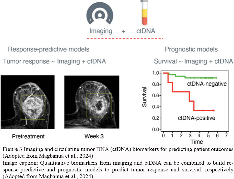 Figure 3 Imaging and circulating tumor DNA (ctDNA) biomarkers for predicting patient outcomes (Adopted from Magbanua et al., 2024) Image caption: Quantitative biomarkers from imaging and ctDNA can be combined to build response-predictive and prognostic models to predict tumor response and survival, respectively (Adopted from Magbanua et al., 2024) |
4.2 Radiation therapy planning and monitoring
Radiation therapy planning and monitoring heavily rely on advanced imaging techniques. CT imaging is essential for delineating the target volume and planning the precise delivery of radiation doses (Forrest, 2016). Positron emission tomography (PET) imaging is increasingly used to visualize and quantify tumor characteristics at a molecular level, which aids in refining radiation therapy plans and assessing treatment response (Unterrainer et al., 2020). PET imaging provides insights into tumor metabolism and receptor expression, which are critical for predicting patterns of failure and adjusting treatment plans accordingly (Marcu et al., 2018). These imaging modalities ensure that radiation therapy is accurately targeted, minimizing damage to surrounding healthy tissues and improving treatment outcomes.
4.3 Assessing surgical success and recurrence
Imaging is indispensable in assessing surgical success and detecting tumor recurrence in veterinary oncology. Pre-surgical imaging, particularly with CT, helps in evaluating the extent of tumors and determining the feasibility of complete resection (Forrest, 2016). Post-surgical imaging is used to confirm the success of the surgery and to monitor for any signs of recurrence. Radiomics, a technique that extracts quantitative features from medical images, is gaining attention for its potential to predict surgical outcomes and recurrence by analyzing pre- and post-treatment images (Shi et al., 2018). This approach allows for a more personalized assessment of surgical success and the likelihood of recurrence, facilitating timely interventions if needed.
In summary, imaging is integral to the monitoring and prediction of treatment outcomes in veterinary oncology. It enhances the precision of chemotherapy and radiation therapy assessments and provides critical insights into surgical success and recurrence, ultimately contributing to more effective and personalized cancer care (Staal et al., 2020).
5 Case Study: Imaging in the Diagnosis and Treatment of a Canine Soft Tissue Sarcoma
5.1 Case introduction
Soft tissue sarcomas (STS) in dogs are locally aggressive tumors that require precise diagnostic and treatment strategies to ensure effective management. These tumors often necessitate surgical resection as the primary treatment modality, with imaging playing a crucial role in both diagnosis and treatment planning. The case presented involves a canine patient diagnosed with a soft tissue sarcoma, highlighting the integration of various imaging techniques to optimize treatment outcomes.
5.2 Imaging techniques used (X-ray, CT, MRI)
In the diagnosis and treatment of canine soft tissue sarcoma, multiple imaging modalities are employed to provide comprehensive insights into the tumor's characteristics and extent. X-rays, while not the primary modality for soft tissue sarcomas, can be useful in initial assessments to rule out bone involvement or detect any calcifications associated with the tumor (Marcello et al., 2015). Computed tomography (CT) offers detailed cross-sectional images that are invaluable for assessing the tumor's size, location, and potential involvement with surrounding structures. This modality is particularly useful in planning surgical approaches and evaluating the feasibility of techniques like MR-guided high-intensity focused ultrasound (HIFU) (Seward et al., 2018). Magnetic resonance imaging (MRI) is considered the gold standard for soft tissue evaluation due to its superior contrast resolution. It provides detailed information on the tumor's margins and its relationship with adjacent tissues, which is critical for surgical planning and assessing the response to neoadjuvant therapies. MRI can also guide the application of advanced techniques such as fluorescence-guided surgery (Spinnato et al., 2021; Beer et al., 2023).
5.3 Outcome and imaging follow-up
The integration of these imaging techniques facilitated a comprehensive treatment plan for the canine patient. Post-surgical imaging follow-up, including MRI, was crucial in assessing the surgical margins and ensuring complete tumor resection. Optical coherence tomography (OCT) was also employed to evaluate surgical margins in real-time, enhancing the accuracy of the resection and reducing the likelihood of local recurrence (Dornbusch et al., 2020). The use of advanced imaging systems, such as fluorescence-based intraoperative imaging, further assisted in distinguishing neoplastic from normal tissue, thereby improving surgical outcomes (Dewitt et al., 2016).
In conclusion, the case underscores the pivotal role of imaging in the diagnosis and treatment of canine soft tissue sarcomas, demonstrating how a multimodal imaging approach can enhance surgical precision and improve patient outcomes.
6 Challenges and Limitations of Imaging in Veterinary Oncology
6.1 High cost of advanced imaging modalities
Advanced imaging techniques such as CT and MRI are crucial for accurate diagnosis and treatment planning in veterinary oncology, particularly for complex cases like head and neck cancers (Hansen and Kent, 2019). However, these modalities are often expensive, which can limit their accessibility and use in veterinary practices. The high cost is a significant barrier, especially in settings where financial resources are constrained, impacting the ability to provide comprehensive care (Forrest, 2016; Klopfleisch and Bauer, 2016).
6.2 Accessibility of imaging equipment
While CT imaging has become more prevalent in veterinary oncology due to its effectiveness in tumor staging and treatment planning, the availability of such equipment is still limited in many regions (Forrest, 2016). This lack of accessibility can hinder timely diagnosis and treatment, particularly in low-income and middle-income areas where imaging resources are scarce. The disparity in equipment availability can lead to unequal care and outcomes for veterinary patients across different geographic locations (Da Silva Froes, 2016).
6.3 Variability in image interpretation
The interpretation of imaging results can vary significantly among practitioners, which poses a challenge in veterinary oncology. Each imaging modality, whether it be ultrasound, x-ray, CT, or MRI, has its own set of advantages and limitations, and the skill level of the interpreter can greatly influence diagnostic accuracy (Klopfleisch and Bauer, 2016). This variability can lead to differences in diagnosis and treatment plans, potentially affecting patient outcomes. Moreover, the complexity of interpreting advanced imaging results requires specialized training, which may not be uniformly available across all veterinary practices (Sotoudeh et al., 2016; Serkova et al., 2020).
In summary, while imaging plays a vital role in the diagnosis and treatment of veterinary oncology, challenges such as high costs, limited accessibility, and variability in interpretation can impede its effectiveness. Addressing these issues is crucial for improving the quality and consistency of care in veterinary oncology.
7 Future Directions in Veterinary Oncology Imaging
7.1 Advancements in imaging technology
The field of veterinary oncology imaging is poised for significant advancements with the continuous development of imaging technologies. Advanced imaging modalities such as CT and MRI are already enhancing the precision of diagnosis and treatment planning by providing detailed anatomical information, particularly for complex cases like head and neck cancers (Forrest, 2016; Hansen and Kent, 2019). The integration of molecular imaging techniques, such as PET, is also on the horizon, offering the potential for more personalized cancer therapy by allowing in vivo visualization of molecular pathways and tumor characteristics (Marcu et al., 2018). These advancements are expected to improve the accuracy of tumor staging and the effectiveness of treatment plans.
7.2 Role of artificial intelligence in imaging analysis
Artificial Intelligence (AI) is set to revolutionize imaging analysis in veterinary oncology by enhancing the interpretation of complex imaging data. AI algorithms can assist in the detection and characterization of tumors, potentially increasing the sensitivity and specificity of imaging modalities like CT and MRI (Forrest, 2016; Strohmayer and Ansón, 2018). The application of AI in imaging could lead to more accurate and faster diagnosis, improved treatment planning, and better monitoring of treatment responses. As AI technology continues to evolve, its integration into veterinary oncology imaging will likely become more prevalent, offering new insights and efficiencies in cancer care.
7.3 Global access and cost reduction strategies
Ensuring global access to advanced imaging technologies and reducing associated costs are critical future directions in veterinary oncology. While CT and MRI are becoming more available, their high costs can limit accessibility, particularly in resource-constrained settings (Da Silva and Froes, 2016; Forrest, 2016). Strategies to reduce costs and improve access include the development of more affordable imaging equipment, training programs to increase the number of skilled operators, and the implementation of telemedicine solutions to share expertise across regions. By addressing these challenges, the benefits of advanced imaging can be extended to a broader range of veterinary patients worldwide.
In summary, the future of veterinary oncology imaging is bright, with advancements in technology, AI integration, and strategies to improve global access and affordability. These developments promise to enhance the diagnosis, treatment, and overall management of cancer in veterinary patients, ultimately improving outcomes and quality of life.
8 Concluding Remarks
Imaging plays a pivotal role in the field of veterinary oncology, serving as a cornerstone for diagnosis, staging, and treatment planning. Techniques such as computed tomography (CT) and magnetic resonance imaging (MRI) are essential for defining the extent of disease, particularly in complex cases like head and neck cancers. These advanced imaging modalities provide detailed anatomical information that aids in surgical and radiation therapy planning, ensuring precise treatment delivery. Despite the availability of advanced imaging, traditional methods like radiography and ultrasonography remain fundamental tools for initial screening and diagnosis, offering a cost-effective and accessible option for many veterinary practices.
The integration of imaging in veterinary oncology significantly enhances diagnostic accuracy and informs treatment decisions. CT imaging, for instance, is crucial for tumor staging and presurgical evaluation, guiding the extent of surgical resection and the feasibility of complete tumor removal. Moreover, imaging modalities like PET are advancing personalized cancer therapy by allowing in vivo visualization of molecular pathways, which can lead to more targeted and effective treatments. These imaging techniques not only improve the precision of treatment but also contribute to better prognostic assessments by enabling continuous monitoring of treatment response and disease progression.
The future of veterinary oncology imaging is promising, with ongoing advancements in technology and methodology. The development of novel imaging techniques and radiotracers, particularly in the realm of molecular imaging, is expected to further personalize cancer treatment and improve outcomes. As these technologies become more widely available, they will likely transform the landscape of veterinary oncology, offering more comprehensive diagnostic and therapeutic options for cancer patients. Continued research and innovation in imaging will be crucial in enhancing the quality of care and extending the lives of veterinary patients.
Acknowledgments
I express our heartfelt gratitude to the two anonymous reviewers for their valuable comments on the manuscript.
Conflict of Interest Disclosure
The author affirms that this research was conducted without any commercial or financial relationships that could be construed as a potential conflict of interest.
Beer P., Pauli C., Haberecker M., Grest P., Beebe E., Fuchs D., Markkanen E., Krudewig C., and Nolff M., 2023, Cross-species evaluation of fibroblast activation protein alpha as potential imaging target for soft tissue sarcoma: a comparative immunohistochemical study in humans, dogs, and cats, Frontiers in Oncology, 13: 1210004.
https://doi.org/10.3389/fonc.2023.1210004
PMid:37727209 PMCid:PMC10505752
Da Silva D., and Froes T., 2016, Veterinary oncology: radiology and ultrasonography exams in decision makingOncologia veterinária – exames radiográficos e ultrassonografia na tomada de decisões, Clínica Veterinária, 124: 56-76.
https://doi.org/10.46958/rcv.2016.xxi.n.124.p.56-76
Derlin T., Grünwald V., Steinbach J., Wester H., and Ross T., 2018, Molecular imaging in oncology using positron emission tomography, Deutsches Arzteblatt international, 115(11): 175-181.
https://doi.org/10.3238/arztebl.2018.0175
Dewitt S., Eward W., Eward C., Lazarides A., Whitley M., Ferrer J., Brigman B., Kirsch D., and Berg J., 2016, A novel imaging system distinguishes neoplastic from normal tissue during resection of soft tissue sarcomas and mast cell tumors in dogs, Veterinary Surgery: VS, 45(6): 715-722.
https://doi.org/10.1111/vsu.12487
PMid:27281113 PMCid:PMC4970890
Dornbusch J., Selmic L., Huang P., Samuelson J., McLaughlin E., Wavreille V., Ogden J., Abrams B., Kalamaras A., Green E., Hostnik E., Every L., Fuerst J., Jennings R., Premanandan C., Lorbach J., Linn S., Alex A., Sorrells J., Yang L., and Boppart S., 2020, Diagnostic accuracy of optical coherence tomography for assessing surgical margins of canine soft tissue sarcomas in observers of different specialties, Veterinary surgery : VS, 50(1): 111-120.
https://doi.org/10.1111/vsu.13510
Forrest L., 2016, Computed tomography imaging in oncology, the veterinary clinics of North America, Small Animal Practice, 46(3): 499-513.
https://doi.org/10.1016/j.cvsm.2015.12.007
PMid:26851976
Freeman L., 2013, Imaging in veterinary oncology: exciting new developments, Veterinary Journal, 197(3) 523-524.
https://doi.org/10.1016/j.tvjl.2013.06.018
PMid:23886702
Hansen K., and Kent M., 2019, Imaging in non-neurologic oncologic treatment planning of the head and neck, Frontiers in Veterinary Science, 6: 90.
https://doi.org/10.3389/fvets.2019.00090
PMid:30984771 PMCid:PMC6448413
Klopfleisch R., and Bauer N., 2016, Basic principles of cancer diagnostics, Cham: Springer International Publishing, 2016: 19-36.
https://doi.org/10.1007/978-3-319-41124-8_2
Lawrence J., Rohren E., Provenzale J., and Provenzale J., 2010, PET/CT today and tomorrow in veterinary cancer diagnosis and monitoring: fundamentals, early results and future perspectives, Veterinary and Comparative Oncology, 8(3): 163-187.
https://doi.org/10.1111/j.1476-5829.2010.00218.x
PMid:20691025
Magbanua M., Li W., and Van ’t Veer L., 2024, Integrating imaging and circulating tumor DNA features for predicting patient outcomes, Cancers, 16(10): 1879.
https://doi.org/10.3390/cancers16101879
PMid:38791958 PMCid:PMC11120531
Marcello B., Gieger T., Jiménez D., and Granger L., 2015, Detection of comorbidities and synchronous primary tumours via thoracic radiography and abdominal ultrasonography and their influence on treatment outcome in dogs with soft tissue sarcomas, primary brain tumours and intranasal tumours, Veterinary and Comparative Oncology, 13(4): 433-442.
https://doi.org/10.1111/vco.12063
PMid:23968175
Marcu L., Moghaddasi L., and Bezak E., 2018, Imaging of tumor characteristics and molecular pathways with pet: developments over the last decade toward personalized cancer therapy, International Journal of Radiation Oncology, Biology, Physics, 102(4): 1165-1182.
https://doi.org/10.1016/j.ijrobp.2018.04.055
PMid:29907486
Mattoon J., and Bryan J., 2013, The future of imaging in veterinary oncology: learning from human medicine, Veterinary Journal, 197(3): 541-552.
https://doi.org/10.1016/j.tvjl.2013.05.022
PMid:23810184
Randall E., 2016, PET-computed tomography in veterinary medicine, the veterinary clinics of North America, Small Animal Practice, 46(3) 515-533.
https://doi.org/10.1016/j.cvsm.2015.12.008
PMid:27068445
Reetz J., 2016, Advanced diagnostic imaging in small animal oncology, Medicine, 2016: 52234196.
Seiler S., Baumgartner C., Hirschberger J., Beer A., Brühschwein A., Kreutzmann N., Laberke S., Wergin M., Meyer-Lindenberg A., Brandl J., Von Thaden A., Farrell E., and Schwaiger M., 2015, Comparative oncology: evaluation of 2-Deoxy-2-[18F]fluoro-D-glucose (FDG) positron emission tomography/computed tomography (PET/CT) for the staging of dogs with malignant tumors, PLoS ONE, 10(6): e0127800.
https://doi.org/10.1371/journal.pone.0127800
PMid:26068641 PMCid:PMC4466332
Serkova N., Glunde K., Haney C., Farhoud M., De Lille A., Redente E., Simberg D., Westerly D., Griffin L., and Mason R., 2020, Preclinical applications of multi-platform imaging in animal models of cancer, Cancer Research, 81: 1189-1200.
https://doi.org/10.1158/0008-5472.CAN-20-0373
PMid:33262127 PMCid:PMC8026542
Seward M., Daniel G., Ruth J., Dervisis N., Partanen A., and Yarmolenko P., 2018, Feasibility of targeting canine soft tissue sarcoma with MR-guided high-intensity focused ultrasound, International Journal of Hyperthermia, 35: 205-215.
https://doi.org/10.1080/02656736.2018.1489072
PMid:30303425
Shi L., He Y., Yuan Z., Benedict S., Valicenti R., Qiu J., and Rong Y., 2018, Radiomics for response and outcome assessment for non-small cell lung cancer, Technology in Cancer Research & Treatment, 17: 1533033818782788.
https://doi.org/10.1177/1533033818782788
PMid:29940810 PMCid:PMC6048673
Sotoudeh H., Sharma A., Fowler K., McConathy J., and Dehdashti F., 2016, Clinical application of PET/MRI in oncology, Journal of Magnetic Resonance Imaging, 44(2): 265-276.
https://doi.org/10.1002/jmri.25161
PMid:27007987
Spinnato P., Kind M., Loarer L., Bianchi G., Colangeli M., Sambri A., Ponti F., Van Langevelde K., and Crombé A., 2021, Soft tissue sarcomas: the role of quantitative MRI in treatment response evaluation, Academic Radiology, 29(7): 1065-1084.
https://doi.org/10.1016/j.acra.2021.08.007
Staal F., Van Der Reijd D., Taghavi M., Lambregts D., Beets-Tan R., and Maas M., 2020, Radiomics for the prediction of treatment outcome and survival in patients with colorectal cancer: a systematic review, Clinical Colorectal Cancer, 20(1): 52-71.
https://doi.org/10.1016/j.clcc.2020.11.001
PMid:33349519
Strohmayer C., and Ansón A., 2018, The role of computed tomography in the oncologic patient, Clin. Vet. Peq. Anim, 38 (1): 7-14
Unterrainer M., Unterrainer M., Eze C., Ilhan H., Marschner S., Roengvoraphoj O., Schmidt-Hegemann N., Walter F., Kunz W., Rosenschöld P., Jeraj R., Albert N., Albert N., Grosu A., Niyazi M., Niyazi M., Bartenstein P., Bartenstein P., Belka C., and Belka C., 2020, Recent advances of PET imaging in clinical radiation oncology, Radiation Oncology (London, England), 15: 1-15.
https://doi.org/10.1186/s13014-020-01519-1
PMid:32317029 PMCid:PMC7171749


. PDF(0KB)
. HTML
Associated material
. Readers' comments
Other articles by authors
. Jianli Zhong
Related articles
. Veterinary oncology
. Imaging techniques
. Tumor staging
. Treatment monitoring
. Artificial intelligence
Tools
. Email to a friend
. Post a comment