2. Faculty of Veterinary Science, University of Agriculture, Faisalabad, Pakistan
3. Livestock and Dairy Development Department, Government of Punjab
Faculty of Veterinary Science, University of Agriculture, Faisalabad, Pakistan
Livestock and Dairy Development Department, Government of Punjab
Department of Clinical Medicine & Surgery, University of Agriculture, Faisalabad, Pakistan
Department of Clinical Medicine & Surgery, University of Agriculture, Faisalabad, Pakistan
 Author
  Correspondence author
International Journal of Molecular Veterinary Research, 2013, Vol. 3, No. 1 doi: 10.5376/ijmvr.2013.03.0001
Received: 03 Dec., 2012 Accepted: 29 Jan., 2013 Published: 17 Feb., 2013
Raza et al., 2013, Cherry Eye: Prolapse of Third Eyelid Gland in Dog- A Case Report, International Journal of Molecular Veterinary Research, Vol.3, No.1 1-3 (doi: 10.5376/ ijmvr.2013.03.0001)
Third eyelid covers the medial canthus of the eye and consists of T-shaped flap like cartilage and tear gland, both are helpful in protection of eye. Prolapsed gland appeared as a dark pink to reddish mass and misdiagnosed as a tumor and treated like a tumor in which gland is excised out. The present report describes a case of cherry eye (prolapse of third eyelid) in 18 months old Cocker spaniel. The case was treated by adopting massage method to replace the third eyelid back to its place followed by administration of eye drops. The treatment method was successful as there was no recurrence when the animal was followed-up for 3 months.
Introduction
Cherry eye is a common ophthalmic malady of dogs and rarely of cats in which eversion / prolapse of third eyelid gland does occur. The prolapsed third eyelid makes it vulnerable to the outer environment. Breeds especially Pekingese, Neapolitan Mastiff, Cocker Spaniel, Beagle, Bulldog and Basset Hound are more prone to this pathological syndrome (Herrera, 2005; Moore, 1998). The disease could occur in any age but most common in young ones i.e. puppies. This can occur in 2~3 years of age and may be unilateral or bilateral (Christmas, 1992; Gellat, 1991). Genetic basis of the disease are not identified and third eyelid is important in protection of eyes as well as production of tears (Gellat, 1991). The eversion of nictitating gland is written off as glandular hyperplasia, hypertrophy, nictitating gland adenoma, protrusion of gland or cherry eye (Mitchel, 2012). The main cause of prolapse is weakening of supportive ligament that fixes the gland (Schoofs, 1999). The present manuscript is a maiden attempt to report the cherry eye disease in dogs from Pakistan.
Case Description
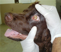
A 1.5 years old male dog (Cocker Spaniel) was presented to Veterinary Teaching Hospital (VTH), Dept. of Clinical Medicine and Surgery, University of Agriculture, Faisalabad, Pakistan, with a complaint of pinkish lump like structure protruding out at the base of left eye from the medial canthus. The size of the structure was similar to that of cherry with bright pink color. This condition was 15 days standing and the patient was in great stress from the last 5 days due to severe irritation and lacrimation (Figure 1).
 Figure 1 Prolapsed third eyelid in dog (Cockerel Spaniel) |
Physical examination revealed that temperature of the animal was normal i.e. 101.6 0F with severe panting and salivation. Other parameters (Respiration 80 bpm and pulse 90 per minute) were also recorded. Regarding previous treatment, patient was treated with eye drops and systemic antibiotics.
The patient was treated by applying the Lignocaine gel on eye (Lidex®, Caraway, Pakistan) and gently massaging the protruded mass clockwise and anti-clock wise by closing the eyelids. After giving 3 rounds of massage each of 4~5 minutes, the prolapsed gland was replaced back to its original position (Figure 2). Then patient was medicated topically with eye drops (Mebradex®, MediPak, Pakistan) to keep the eye surface wet and reduce the chances of inflammation and infection. The animal was monitored for recurrence and there was no recurrence up to 3 months post treatment.
 Figure 2 Reduced prolapse of third eyelid after application of massage method |
Discussion
Third eyelid covers the medial canthus of the eye, consists of T-shaped flap like cartilage and tear gland, both are helpful in protection of eye (2). Prolapsed gland appeared as a dark pink to reddish mass and misdiagnosed as a tumor and treated like a tumor in which gland was excised out, but this resulted in dryness of the eye because third eyelid gland or nictitating gland is one of the tear producing glands that keeps the eye moist. The main complication after its removal was kerato-conjunctivitis siccas (KCS) (Gelatt, 1999). Third eyelid gland produces 30% of the total tears (Gellat, 1991; Saito et al., 2001) which are important for the intactness of eyelid, eyeball surface and conjunctiva (Davidson and Kuonen, 2004). This prolapse happens because of the loss of tensile strength of the peri-orbital supporting ligament that anchors the gland to the peri-orbit (Mitchel, 2012). So the prolapsed gland becomes exposed to the external environment which leads to increase in the glandular size due to abrasion and drying (Moore, 1998; Gellat, 1991).
Regarding its treatment, two methods are usually adopted; excision of gland and replacement of gland. Excision of gland is an old method and not recommended now-a-days because the whole gland is nipped at its base which leads to 'dry eye’. This causes further complications. Regarding second option, cosmetically correction of prolapsed gland is the most recommended method in which 'tucking' technique is usually used. Previously single tucking technique was used but if somehow suture may adhere, this will cause blepharospasm and visibility of the suture. So this method is modified now and a wedge of tissue is removed but how much tissue is removed and tiny sutures will tightens the gap or not, are the major points of consideration. Main complications of modified techniques are inflammation, chances of recurrence and failure of stitch holding capacity (http://marvistavet.com/html/cherry_eye.html). The present case was treated by simple massage method followed by no recurrence. So it is suggested that the massage method to replace the prolapsed third eyelid is considered one of the best and safest methods to treat the cherry eye condition in dogs if there is no recurrence.
References
Christmas R.E., 1992, Common ocular problems of Shin Tzu dogs, The Canadian Veterinary Journal, 33(6): 390-393 PMid:17424020 PMCid:1481255
Davidson H.J., and Kuonen V.J., 2004, The tear film and ocular mucins, Vet. Ophth, 7(2): 71-77 http://dx.doi.org/10.1111/j.1463-5224.2004.00325.x PMid:14982585
Gelatt K.N., 1999, Veterinary ophthalmology, 3rd Edition, Lippincott Williams & Wilkins, Philadelphia, pp.1544
Gellat K.N., 1991, Canine lacrimal and nasolacrimal diseases, In: Veterinary Ophthalmology, 2nd Ed., Lea & Febiger, Philadelphia, pp.276-289
Herrera D., 2005, Surgery of the Eyelids, Proceedings of the World Small Animal Veterinary Association Mexico City, Mexico, 1-4 PMid:16203008
Mitchel N., 2012, Third eye lid protrusions in dogs and cats, Veterinary Ireland Journal, 2(4): 205-209
Moore C.P., 1998, Terceira pálpebra. In: Slatter D., Manual de cirurgia de pequenos animais, 2nd Ed., Paulo: Manole, 2: 1428-1435
Saito A., Izumisawa Y., Yamashita K., and Kotani T., 2001, The effect of third eyelid gland removal on the ocular surface of dogs, Vet. Ophth., 4(1): 13-18 http://dx.doi.org/10.1046/j.1463-5224.2001.00122.x
Schoofs S.H., 1999, Prolapse of the gland of the third eyelid in a cat: a case report and literature review, J. American Ani. Hosp. Assoc., 35(3): 240-242 PMid:10333264
. PDF(94KB)
. FPDF(win)
. HTML
. Online fPDF
Associated material
. Readers' comments
Other articles by authors
. A. Raza
. A. Naeem
. M. Ahmad
. A. Manzoor
. M. Ijaz
Related articles
. Dog
. Cherry eye
. Third eyelid prolapse
. Treatment
. Massage method
Tools
. Email to a friend
. Post a comment


