1 Introduction
Pteropus giganteus giganteus presents a seasonally monoestrous and polygynous pattern of reproduction (Marshall, 1947). The reproductive cycle of Pteropus giganteus giganteus exhibits mainly the five stages in their sex cycle. A period of sexual quiescence - from July to August, Oestrus and Fertilisation-from last week of August to first week of September, Preganacy-from Mid September to late February or first week of March, Parturation - During the first week of March and Lactation - from first week of March to July. This bat has gestation period of 140-150 days. The different strategies had been developed for successful reproduction in many species of bats. These were delayed ovulation in Plecotus townsendii, delayed implantation in Fischer’s pygmy fruit bat, Haplonycteris ficheri (Heidmann, 1989) and sperm storage in Pipistrellus kuhlii (Sharifi et al., 2004). Knowledge of reproductive asymmetry and unilateral pregnancy in Chiroptera is due to Wimsatt (1979). Megabats are strictly nocturnal; the only exceptions had been reported were Samoan flying fox (Pteropus samoensis) and the Trogen fruit bat (Pteropus tonganus) which were active during day and night (Utzurrum, 2002). Pteropus giganteus giganteus leave their roost for foraging at dusk and continue feeding until prior to the dawn that is showing the nocturnal habit, so most active during the night hours (Kunz and Diaz, 1995). Out of 18 families of living bats (Hill and Smith, 1988) eight families had been reported in India. The megabats feed mainly on fruit, flowers, nectar and pollen. Thus megabats play a major role in pollination and seed dispersal (Walker and Molur, 2003; Stephenraj et al., 2010; Ul-Hassan et al., 2010). These bats are vital to the survival of rainforests and play crucial role in rejuvenation of the entire ecosystem (Cox et al., 1992). Thus, the Angiosperm's biodiversity of forest can be directly correlated with the population of bats. Due to such ecological and environmental requirement of this megabat, management and conservation needs a special attention. Unfortunately little is known regarding its reproductive aspects, which may be needed for management and conservation in future.Ovaries play crucial role in reproductive physiology due to its role in oogenesis and synthesis of steroid and peptide hormones. Thus the aim of this work is to understand the reproductive biology of Pteropus giganteus giganteus by studyingthe histology and morphometrical changes in ovary during various reproductive phases.
2 Material and Methods
2.1 Collection of Specimens
Pteropus giganteus giganteus (Brunnich) is an Indian megachiropteran bat of pteropidae family commonly known as Indian flying fox. A colony of 50 to 500 individuals had been observed on the large tree of Ficus bengalensis near the water reservoir at Padmapur village. All the specimens used during entire study period were obtained from natural populations from feeding site at Padmapur village near Armori. [Longitude 20°22' North and latitude 79°48' East (Dist-Chandrapur, Maharashtra)]. The specimens were collected from December 2008 up to December 2010 in such a way that entire reproductive cycle was represented. Feeding sites were identified by the examination of guano of the Pteropus giganteus giganteus. Five specimens were collected during each reproductive stage. Complete reproductive cycle of Pteropus giganteus giganteus had been studied and anoestrous, oestrous, early pregnancy, mid pregnancy, parturation and lactation stages were confirmed by the histological examination of uterus and ovaries, morphological examination of mammary glands and breeding behavior at roosting site.
2.2 Morphometric analysis
Mature live animals were brought to the laboratory and anesthetized with ether and killed by decapitation. The ovaries were fixed in alcoholic Bouin’s fixative solution and serially sectioned (thickness-5µm). Ovarian sections were stained with hematoxylin-eosin (Humason, 1979). Histological sections of right and left ovaries were used to calculate the diameter of the ovary. The ovaries and corpus luteum were assumed to be spherical. Ovarian and corpus luteum diameter was measured (a) and the diameter at right angles to this (b) (Wiliams, 1977). The diameter (D) was calculated using the following equation:
Diameter (D) (mm) = .png) ab
ab
The mean diameter (D) for each ovary and corpus luteum was calculated. This was then converted to an ovarian and corpus luteum volume and surface area using the following equations:
Volume of ovary/Corpus luteum (mm3) = 
Surface area of ovary/Corpus luteum (mm2) = 4πr2
2.3 Statistical analysis
Statistical analysis was performed to determine the significant changes in ovarian diameter, surface area and volume of the ovary during the different phases of the reproductive cycle. Mean, Standard error, Standard deviation, Variance and ANOVA with post-hoc Tukey HSD test were calculated using Statistical Package for Social Sciences (SPSS 10.0).
3 Results
3.1 Ovarian morphometric analysis
Non-significant increase in diameter, surface area and volume of both the ovaries has been observed during lactation to anaoestrous and anaoestrous to prooestrous (Table 1 and Table 2). However significant increase in diameter, surface area and volume of both the ovaries has been observed during prooestrous to oestrous (Table 1 and Table 2). During pregnancy one of the ovaries shows corpus luteum, indicating that ovulation has been occurred in this ovary and another undergoes regression. Thus out of six reproductive asymmetry patterns reported in chiroptera, Pteropus giganteus giganteus exhibited a pteropid pattern of the reproductive asymmetry. Non-significant increase in diameter, surface area and volume has been observed in ovary from which ovulation has been occurred during oestrous to early pregnancy (Table 1). From early pregnancy to mid-pregnancy, this ovary showed non significant increase in diameter whereas significant (P<0.05) increase in surface area and volume (Table 3). At the termination of pregnancy and start of lactation significant decrease in diameter, surface area and volume has been reported in the ovary from which ovulation has been occurred (Table1). However, another ovary, referred as regressed ovary, showed significant decrease in diameter, surface area and volume from oestrous to early pregnancy (Table 2). As the pregnancy proceeds, significant decrease in diameter has been observed during mid-pregnancy in regressed ovary. Non significant decrease in surface area and volume has been noted in regressed ovary from early pregnancy to mid pregnancy (Table 3). At the termination of pregnancy and start of lactation significant increase in diameter, surface area and volume has been reported in the regressed ovary (Table 2). Significant increase in diameter, surface area and volume of corpus luteum has also been reported during early pregnancy to mid pregnancy (Table 3).
.png)
Table 1 Comparison of diameter, surface area and volume of right ovary in Pteropus giganteus giganteus during different stages of life cycle
|
.png)
Table 2 Comparison of diameter, surface area and volume of left ovary in Pteropus giganteus giganteus during different stages of life cycle |
.png)
Table 3 Comparison of ovarian diameter in Pteropus giganteus giganteus during pregnancy |
3.2 Histological changes in ovary
3.2.1 Development of Graffian follicle
Development of graffian follicle starts from primordial follicle. Primordial follicles were composed of an immature oocyte surrounded by a single layer of flattened granulosa cells (Figure 1).
.png)
Figure 1 Primordial follicle, hematoxylin-eosin stain. (PRF- Primordial follicle) |
Primary follicles consist of an oocyte surrounded by one to two layers of cuboidal granulosa cells (Figure 2 and Figure 3).
.png)
Figure 2 Primary follicle, hematoxylin-eosin stain. (N-Nucleus; O-Oocyte; PF-Primary follicle) |
.png)
Figure 3 Primary follicle, hematoxylin-eosin stain. (GC-Granulosa cell; N-Nucleus; O-Oocyte; PF-Primary follicle) |
Primary follicles develop into secondary follicles referred as small preantral follicle, which comprised of two to four layers of granulosa cells (Figure 4). Graffian follicles which possess more than two granulosa layers, could be termed as multilaminar follicles.
.png)
Figure 4 Bilaminar Secondary follicle, hematoxylin-eosin stain. (GC-Granulosa cell; SF-Secondary follicle; VM-Vitelline Membrane) |
In successive development small preantral follicle becomes large preantral follicle that had four to six to layers of cuboidal granulosa cells. In large preantral follicles, ovum was surrounded by zona pellucida and four to six layers of granulosa cells (Figure 5).
.png)
Figure 5 Multilaminar follicle, hematoxylin-eosin stain. (N-Nucleus; O-Oocyte; PAF-Preantral follicle; VM-Vitelline Membrane; ZP-Zona pellucida) |
Development of large preantral follicle results in the formation of vesicular follicle (Figure 6).Vesicular follicle comprised of vesicular spaces that appeared in the closely packed cuboidal granulosa cells. Granulosa cells start to congregate towards one side to form the antral space and antrum start to develop in the follicle. Thus graffian follicle was refered as antral follicle (Figure 7). Antral follicle was characterized by the development of follicular antrum and more than five layers of cuboidal granulosa cells. Oocyte was surrounded by thick zona pellucida. The oocyte was pushed to one side of the follicle by developing antrum and separated by two or three layers of cumulus cells. Ovum is attached to the granulosa cells only at one site forming the cumulus oophorus. Cells of carona radiata were in contact of follicular antrum. Thecal layer was present towards the outer side of basement membrane, which was composed of 3-4 layers of cells. Theca layer could be distinguished into theca externa and theca interna (Figure 8).
.png)
Figure 6 Vesicular follicle showing vesicular spaces, hematoxylin- eosin stain. (N-Nucleus; O-Oocyte; VF-Vesicular follicle; VS-Vesicular space; VM-Vitelline Membrane) |
.png)
Figure 7 Antral follicle showing antral cavity, hematoxylin-eosin stain. (FA-Follicular antrum; GC-Granulosa cell; O-Oocyte) |
.png)
Figure 8 Part of antral follicle showing cumulus oophorus. The primary oocyte is attached to zona granulosa by cumulus oophorus, hematoxylin-eosin stain. (CR-Carona radiata; CO-Cumulus oophorus; FA-Follicular antrum; O-Oocyte; VM-Vitelline Membrane; ZP-Zona pellucida) |
As follicle attains maturity oocyte appears to lie in free antral cavity (Figure 9 and Figure 10).
.png)
Figure 9 Preovulatory Graafian follicle. The primary oocyte floats freely within the follicular antrum, hematoxylin-eosin stain. (CR-Carona radiata; FA-Follicular antrum; GC-Granulosa cell; O-Oocyte; VM-Vitelline Menbrane; ZP-Zona pellucida) |
.png)
Figure 10 Preovulatory Graafian follicle. hematoxylin-eosin stain. (CR-Carona radiata; FA-Follicular antrum; GC-Granulosa cell; O-Oocyte) |
All graffian follicles do not attain the maturity. Some of these follicles undergo the phenomenon of atresia. Graffian follicles undergo process of atresia by nuclear pyknosis and nuclear karyorrhexis (Figure 11 and Figure 12).
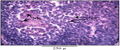
Figure 11 Multilaminar follicle showing atresia. The nuclear pyknosis and nuclear karyorrhexis is clearly visible, hematoxylin-eosin stain. (NK-Nuclear karyorrhexis; NP-Nulear pyknosis) |
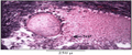
Figure 12 Antral follicle showing atresia, hematoxylin-eosin stain. (NP-Nuclear pyknosis) |
3.2.2 Histoarchitectural changes in the ovary
During anoestrous phase both ovaries showed peripheral cortex having cluster of primordial follicles as well as several follicles in the various stages of maturation; with primordial, primary, secondary, preantral and antral follicles were observed in both the ovaries (Figure 13). Few atretic follicles have been also reported.

Figure 13 Transverse section of ovary during anoestrous phase, hematoxylin-eosin stain. (AF-Atretic follicle; PF-Primary follicles; PRF-Primordial follicle; TA-Tunica albuginea; ZP-Zona pellucida) |
During proestrous both the ovaries become active and ovarian cortex showed numerous primordial follicles as well as primary, secondary, vesicular and tertiary follicle with developing follicular antrum (Figure 14). Increased number of bilaminar and multilaminar follicles were reported.
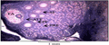
Figure 14 Transverse section of ovary during proestrous phase, hematoxylin-eosin stain. (FA-Follicular antrum; PF-Primary follicles; PRF-Primordial follicle; SF-Secondary follicle ATF-Antral graffian follicle) |
During oestrous both the ovaries show similar pattern (Figure 15). Abundance of all types of follicles in various stages of development was observed. Characteristically primordial follicles and primary oocyte were congregated at periphery of the cortex and intermingled with stromal cells. As the follicles matured from primary to antral state, they appeared to migrate from more cortical positions towards medulla. Larger antral follicles lie close to the periphery of the ovary. Very early antral and atretic follicles were reported in both the ovaries.

Figure 15 Transverse section of ovary during oestrous phase, hematoxylin-eosin stain. (AF-Atretic follicle; ATF-Antral follicle) |
During pregnancy ovary showed presence few bilaminar, multilaminar and other follicles in the stage of atresia. Within the ovary from where ovulation occurs showed presence of single extrovert type corpus luteum which persists up to mid pregnancy and then regresses. Significant increase in size of corpus luteum occurs from early to mid pregnancy. At mid pregnancy corpus luteum occupies 2/3rd portion of the ovary (Figure 16, Figure 17 and Figure 18). During early pregnancy corpus luteum appeared active and secretory. The luteal cells were relatively large with rounded nucleoli and abundant eosinophillic cytoplasm. The entire gland appeared to be well supplied with blood vessels (Figure 19 and Figure 20). By late pregnancy the luteal cells appeared smaller with smaller nucleoli and the corpus luteum becomes infiltered with connective tissue and leucocytes. Another ovary undergoes the regressed stage. In regressed ovary the atretic follicles can be seen and only primordial follicles can be observed after conceiving the pregnancy and in later stages ovary undergoes complete regression showing only cluster of primordial follicles within the ovary (Figure 21).

Figure 16 Transverse section of ovary during early pregnancy showing extrovert type of corpus luteum, hematoxylin-eosin stain. (AF-Atretic follicle; CL-Corpus luteum) |
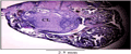
Figure 17 Transverse section of ovary of during mid pregnancy. 70% of the area of the ovary is occupied by the extrovert type of corpus luteum, hematoxylin-eosin stain. (CL-Corpus luteum) |

Figure 18 Extrovert Corpus luteum, hematoxylin-eosin stain. (CL-Corpus luteum) |

Figure 19 Magnified portion of corpus luteum during early pregnancy showing large and small luteal cells, hematoxylin-eosin stain. (BV-Blood vessel; LLC-Large luteal cell; SLC-Small luteal cell) |
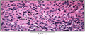
Figure 20 Magnified portion of corpus luteum during mid pregnancy showing large and small luteal cells, hematoxylin-eosin stain. (BV-Blood vessel; LLC-Large luteal cell; SLC-Small luteal cell) |
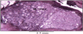
Figure 21 Transverse section of regressed ovary during pregnancy, hematoxylin-eosin stain. (PRF-Primordial follicle) |
During lactation both the ovaries were similar in size. Histologically ovarian cortex showed numerous primordial follicles intermingled with stroma towards periphery. Primordial, unilaminar, bilaminar and few multilaminar follicles were observed in both the ovaries but none of the ovaries showed antral or mature follicle (Figure 22).
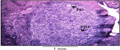
Figure 22 Transverse section of ovary during lactation, hematoxylin-eosin stain. (PR-Primary follicle; PRF-Primordial follicle) |
4 Discussion
Development of graffian follicle in Pteropus giganteus giganteus exhibits mainly five stages of development (Type 1 to type 5). Gopalkrishna et al. (1974) and Krishna and Dominic (1980) had observed the development of garffian follicles in bat Scotophilus heathi. Lundy et al. (1999) had classified the ovine graffian follicles on the basis of morphometric characteristics. Comparing the results of the study of development of graffian follicle, it can be concluded that process of development of graffian follicle in Pteropus giganteus giganteus has similarity with other mammalian species. Histology of ovary had been studied in various species of Indian bats viz. Cynopterus sphinx, Rhinopoma kinneri, Megaderma lyra lyra, Hipposideros bicolor (Gopalkrishna and Moghe, 1960). Rasweiler (1988) had noted the interstitial gland cells in the ovary of Molossus ater which were concerned with the secretion of progesterone. Reproductive asymmetry had been described by Wimsatt (1979) and explained the complete sinistral dominance of ovary in megaderma pattern, contra lateral dominance in miniopterus pattern, dextral dominance in molossid pattern, strong tendancy for right and left ovaries to ovulate alternately in successive cycle in phyllostomid pattern, ovulation may occurs from either ovary with nearly equal frequency in myotis pattern and non random alternation of ovulations between right and left ovaries in successive cycles in pteropid pattern. In Pteropus giganteus giganteus neither ovary is dominant. Both the ovaries contain a variety of follicular types and ovulation can occur from any of these ovaries. Only single ovary is functional during each pregnancy cycle and other undergoes regression. Such pteropid pattern has been observed in Pteropus giganteus giganteus. Marshall (1947, 1949, 1953) in Pteropus giganteus, Ramkrishna (1950) in Cynopterus sphinx, Gopalkrishna (1964, 1969) and Gopalkrishna and Choudhary (1977) in Rousettus leschenaulti, Gopalkrishna and Murthy (1960) in Taphozous longimanus, Matthews (1941) in Nycteris luteola and Nycteris hispida, Rasweiler (1972) in Glossophaga soricina, de Bonilla and Rasweiler (1974) in Carollia perspicillata, Quintero and Rasweiler (1974) in Desmondus rotundus, Rasweiler (1977) in Noctilio albiventris had reported alternation of ovulation between ovaries in successive pregnancy which is similar to that found in Pteropus giganteus giganteus. After ovulation, the corpus luteum was formed from the cellular components of the ovarian follicle, the granulosa and theca cells (Rodgers et al., 1983). The granulosa cells do not divide after ovulation, but they increase in size and undergo morphological changes (Auletta and Flint, 1988). Corpus luteum consists of two types of luteal cells small and large luteal cells. This finding is in agreement with the findings of Christensen and Gillim (1969) and Niswender et al. (1985). The large luteal cells are derived from the granulosa cells while small luteal cells are derived from thecal cells (Rodgers et al., 1983). Gopalkrishna and Badwaik (1998) had studied the growth of the corpus luteum in relation to gestation in Rousettus leschenaulti, Rinopoma microphyllum (kinneri), Megaderma lyra lyra, Rhinoplophus rouxi, Hipposideros fulvus fulvus, Hipposideros speoris and Pipistrellus ceylonicus chrysothrix and reported the intraovarian corpus luteum in which corpus luteum remains within ovary and progressively enlarges in R. leschenaulti and Pipistrellus ceylonicus chrysothrix, pedunculated corpus luteum in Rhinoplophus rouxi and extrovert corpus luteum in Megaderma lyra lyra, Hipposideros fulvus fulvus and H. speoris. Heideman (1989) had observed the corpus luteum in the Fischer’s pygmy fruit bat Haplonycteris fischeri and revealed that corpus luteum was always ipsilateral to reproductive duct carrying the newly ovulated ovum conceptus. Anand kumar (1965) in Rhinopoma kinneri, Gopalkrishna and Bhatia (1983) in Hipposideros speoris, Sapkal and Bhandarkar (1984) and Seraphim (2002) in Hipposideros lankadiva had observed the extrovert type of corpus luteum. Only single extrovert type of corpus luteum has been observed in Pteropus giganteus giganteus. Corpus luteum persists till mid pregnancy and now placenta takes over the function of production of progesterone to maintain successful pregnancy thus corpus luteum gets regressed.
The results of morphometric study of ovaries presents no morphostructural difference between the left and right ovary. Rani and Devi (2011) in human, Flamini et al. (2009) in viscacha, Belloa et al. (2012) in African zebu cattle and Ofusori (2011) in African tree pangolin have noted the morphometric changes in the ovaries. Morphometric analysis does not reflect any significant difference when the data were compared. Hence both the ovary could be the principal sex organ of reproduction in Pteropus giganteus giganteus. Thus results demonstrate that the ovulation in the left ovary and the right ovary is alternate and none of the ovary showed dominance over other like in other pteropid bats.
Auletta F.J., and Flint A.P., 1988, Mechanisms controlling corpus luteum function in sheep, cows, nonhuman primates and women especially in relation to the time of luteolysis, Endocrine Reviews, 9:88-105
http://dx.doi.org/10.1210/edrv-9-1-88 PMid:3286237
Belloa A., Adamub Y.A., Umaruc M.A., Garbad S., Abdullahid A.U., Adamud M.K., Saidue B., Ukashatuf S., Henaa S.A., and Mahmudag A., 2012, Morphometric analysis of the reproductive system of African zebu cattle, Scientific Journal of Zoology, 1(2): 31-36
Cox P.A., Elmiqvist T., Pierson E.D., and Rainey W.E., 1992, Flying foxes as pollinaters and seed dispersers in Pacific Island Ecosystems, In : Pacific Island Flying Foxes : Proceedings of an International Conversation Conference (eds D. E. Wilson and G.L. Graham) pp. 18 – 23 US Fish and Wild life service Biological Report 90 (23). US Department of the Interior, Fish and Wild life service, Washington DC.
Christensen A.K., andGillim S.W.,1969,The correlation of fine structure and function in steroid-secreting cells, with emphasis on those of gonads, In:The Gonads (K. W.McKerns, ed). New York: Appleton-Century-Crofts. pp.415-488
Bonilla H., and Rasweiler J.J., 1974, Breeding activity, pre implantation development and oviduct histology of short tailed fruit bat Carollia, in captivity,The Anatomical Record, 179: 385-403
http://dx.doi.org/10.1002/ar.1091790309 PMid:4837767
Flamini M.A., Barbeito C.G., Gimeno E.J., and Portiansky E.L., 2009, Histology, histochemistry and morphometry of the ovary of the adult plains viscacha (Lagostomus maximus) in different reproductive stages, Acta Zoologica, 90: 390-400
http://dx.doi.org/10.1111/j.1463-6395.2008.00386.x
Gopalkrishna A., Madhavan A., Thakur R.S., and Rajgopal G., 1974, The graffian follicle in some Indian bats, Current Science, 43: 400-401
Gopalkrishna A., and Moghe M.A., 1960, Observations on the ovaries of some Indian bats, Proceedings of the National Institute of the Sciences of India, 26: 11-19
Gopalkrishna A., and Bhatia D., 1983, Breeding habits and associated phenomena in some Indian bats. Part VII-Hipposideros speoris (Schneider) (Hipposideridae) from Chandrapur, Maharashtra, Journal of Bombay Natural History Society, 79: 549-556
Gopalkrishna A., and Badwaik N., 1998, Growth of the corpus luteum in relation to gestation in some Indian bats, Current science, 57(16): 883-886
Gopalkrishna A., and Murthy K.V.R., 1960, Utero-ovarian junction in two species of bats, Bulletin of Zoological society, Nagpur College of Science, 3: 19-22
Gopalkrishna A., 1964, Post-partum pregnancy in the Indian fruit bat, Rousettus leschenaulti (Desmarest), Current Science, 33: 558-559
Gopalkrishna A., 1969, Unusual persistence of the corpus luteum in the Indian fruit bat, Rousettus leschenaulti (Desmarest), Current Science, 38: 388-389
Gopalkrishna A., and Choudhary P.N., 1977, Breeding habits and associated phenomenon in some Indian bats Part I. –Rousettus leschenaultia (Desmaret) – Megachiroptera, Bombay Natural History Society, 74: 1-16
Heideman P.D., 1989, Delayed development in Fischer’s pygmy fruit bat, Haplonycteris fischeri, in the Philippines, Journal of Reproduction and Fertility, 85: 363-382
http://dx.doi.org/10.1530/jrf.0.0850363 PMid:2703981
Hill J.E. and Smith J.D., 1988, Bats, A Natural History British Museum (Natural History), London.
Humason G.L. 1979, Animal tissue techniques. 4th Edn. W.Freeman and Company, SanFrancisco.
Kunz T. H., and Diaz C.A., 1995, Folivary in friit eating bats,with new evidence from Artibeus jamdicenisis (Chiroptera : Phyllostomidae ), Biotropica, 27:106-120
http://dx.doi.org/10.2307/2388908
Krishna A., and Dominic C.J., 1980, Observation on the Graafian follicles of the vespertiliponid bat, Scotophilus heathi, Proceedings of Zoological Society, Calcutta, 33: 1-7
Lundy T., Smith P., O’Connell A., Hudson N.L., and McNatty K.P., 1999, Populations of granulosa cells in small follicles of the sheep ovary, Journal of Reproduction and Fertility, 115: 251-262
http://dx.doi.org/10.1530/jrf.0.1150251 PMid:10434930
Marshall A.J., 1949, Pre-gestational changes in the giant fruit bat ( Pteropus giganteus ), with special reference to an asymmetric endometrial reaction, Proceedings of the Linnean Society of London, 161:26-36
http://dx.doi.org/10.1111/j.1095-8312.1949.tb00534.x
Marshall A.J., 1953, The unilateral endometrial reaction in the giant fruit-bat (Pteropus giganteus Brünnich), Journal Endocrinology, 9: 42-44
http://dx.doi.org/10.1677/joe.0.0090042 PMid:13034980
Matthews L.H., 1941, Notes on the genitalia and reproduction of some African bats, Proceedings of the Zoological Society of London, B 111: 289-346
Niswender G.D., Schwall R.H., Fitz T.A., Farin C.E., and Swyer H.R., 1985, Regulation of luteal function in domestic ruminants, New concept, Rec.Prog. Horm.Res., 41:101-151 PMid:2996087
Ofusori D.A., 2011, The morphostructure of the ovary of African tree Pangolin Manis tricuspis, The Internet Journal of Veterinary Medicine, 4 (1) DOI: 10.5580/ea2v
Quintero F., and Rasweiler J.J., IV 1974, Ovulation and early embryonic development in the captive vampire bat Desmondus rotundus, Journal of Reproduction and Fertility, 41: 265 - 273
http://dx.doi.org/10.1530/jrf.0.0410265 PMid:4452969
Ramkrishna P.A., 1950, Reproduction in Cynopterus sphinx (Vahl.), Proceedings of the National Institute of Sciences, India 16: 362
Rani U.V., and Devi S.V., 2011, Morphological and morphometric parameters of human ovaries from embryonic to menopausal age, International Journal of Applied and Basic Medical Research,1(2) : 89-92
http://dx.doi.org/10.4103/2229-516X.91151 PMid:23776784 PMCid:PMC3657960
Rasweiler J.J., IV 1972, Reproduction in the long-tongued bat, Glossophaga soricina, I. Preimplantation development and histology of the oviduct, Journal of Reproduction and Fertility, 31: 249-262
http://dx.doi.org/10.1530/jrf.0.0310249 PMid:4118051
Rasweiler J.J., IV 1974, Reproduction in the long-tongued bat Glossophaga soricina. II. Implantation and early embryonic development, The American Journal of Anatomy, 139: 1-36
http://dx.doi.org/10.1002/aja.1001390102 PMid:4810014
Rasweiler J.J., IV 1977, Preimplantation development, fate of zona pellucida, and observations on the glycogen rich oviduct of the little bulldog bat, Noctilio albiventris, The American Journal of Anatomy, 150: 269-299
http://dx.doi.org/10.1002/aja.1001500205 PMid:562617
Rodgers R. J., O’Shea J.D., and Findlay J.K., 1983, Progesterone production in vitro by small and large ovine luteal cells, Journal of Reproduction and Fertility, 69:113-124
http://dx.doi.org/10.1530/jrf.0.0690113 PMid:6310106
Sapkal V.M., and Bhandarkar W.R., 1984, Breeding habits and associated phenomena in some Indian bats. Part IX – Hipposideros lankadiva (Kellart)-(Hipposideridae), Journal of Bombay natural history society, 81: 380-386
Stephenraj D., Isaac S.S., Kunz T.H., and Stanley J.A., 2010, Foraging behaviour ofMegachiropteran Bats (Chiroptera: Pteropodidae) in Courtallam, Tamilnadu, South India, Bioinfolet, 7 (2):175-180
Seraphim E. R., 2002, Endocrine interaction during different phages of the female reproductive cycle in Hipposideros lankadiva ( Kelaart ). Thesis submitted to RTM Nagpur University, Nagpur.
Sharifi M., Ghiorbani R., Fazeli A., and Holt W., 2004, Evidence of sperm storage in Pipistrellus kuhlii (Chiroptera : Vespertilionidae ) in western Iran, Folia zoologica, 53 (1) : 1-6.
Ul-Hassan M.M., Gulraiz T.L., Rana S.A., and Javid A., 2010, The diet of Indian flying-foxes (Pteropus giganteus) in urban habitats of Pakistan, Acta Chiropterologica, 12(2):341-347
http://dx.doi.org/10.3161/150811010X537927
Utzurrum R.C., 2002, Natural History guide to American Samoa; A collection of articles (ed. Craig P.), National park of American Samoa Dept, Marine and wildlife Resources, Samoa
Walker S., and Molur M., 2003, Summary of the status of South Indian Asian chiroptera: Conservation assessment and management plan workshop report, Madurai, pp.1-24
Wiliams M., 1977, Quantitative methods in Biology, Amsterdam: Elsevier/North-Holland Biomedical press
Wimsatt W.A., 1979, Reproductive asymmetry and unilateral pregnancy in chiroptera, Journal of Reproduction and Fertility, 56: 345-357
http://dx.doi.org/10.1530/jrf.0.0560345 PMid:38165

 Author
  Correspondence author
.png)
.png)
.png)
.png)
.png)
.png)
.png)
.png)
.png)
.png)
.png)
.png)
.png)
.png)
.png)
.png)
.png)
.png)
.png)
.png)
.png)
.png)


