Histomorphometric study of the prenatal Development of the Circumvallate Papillae of one-humped camel (Camelus dromedarius) 
 Author
  Correspondence author
International Journal of Molecular Zoology, 2015, Vol. 5, No. 1 doi: 10.5376/ijmz.2015.05.0001
Received: 29 Oct., 2014 Accepted: 28 Feb., 2015 Published: 09 Mar., 2015
Bello et al., 2015, Histomorphometric study of the prenatal Development of the Circumvallate Papillae of one-humped camel (Camelus dromedarius), International Journal of Molecular Zoology, Vol.5, No.1 1-5 (doi: 10.5376/ijmz.2015.05.0001)
This study aimed at investigating the prenatal development of camel circumvallate papillae using standard histomorphometric methods. In the experiment, fifteen Camelus dromedarius foetuses obtained from Sokoto metropolitan abattoir at different gestational ages were used for the study. The fetuses were weighed and grouped according to their gestational ages which were estimated using their crown-vertebral-rump length. From the fifteen tongues used for the study, five belonged to the foetuses being in the first trimester of prenatal development, five came from the second trimester and the next five from the third trimester. A total of ten foetuses were males and five were females. In all stages of development, the tongues were elongated, with flattened and rounded apex .At the first trimester, the tongues were almost uniform width and thickness throughout the length. They were uniformly pinkish, withoutpigmentation and no visiblelingual papillae. At the second trimester, the tongues were observed to have taken the normal shape of an adult tongue with variable size and shape of lingual papillae; tapering rostrally from the root to the apex. Biometrically, the weight of the foetuses were found to be 0.18±0.05 to 21.70±7.28 kg from the first to the second trimester, the crown – vertebral – rump – length were found to be 15.75±4.42 to 94.00±2.83cm from the first trimester to the third trimester, weight of the head were found to be 25.05±15.17 to 1120.00±14.14g and weight of the tongues were found to be 0.79±0.22 to 116.25±11.49g from first trimester to third trimester. Histological observations showed that the circumvallate papillae were coveredwith keratinized stratified squamous epithelium, which was composed by basal, spinosum, granulosum and corneum layers. Lingual glands were located in the deeper parts of the papillae. The surface of the epithelium was less keratinised compared to that of the surrounding surface except the peripheral parts of the papilla. Dermal inter-digitations of variable sizes into the epidermis were observed along the whole surface. Few taste buds were observed along the medial papillary wall epithelium of the small-sized papillae.
The one-humped Dromedary (Camelus dromedarius) and the two-humped Bactrian (Camelus bactrianus) camels are among the largest mammalian species, adapted to the desert where thorny plants with rough and hard stems grow and with its high temperatures and extreme desiccation (2, 3). The camel’s mouth is very sturdy and is developed to maintain efficient feeding of these plants and is rubbery so that thorns and branches won’t damage it (2, 4).
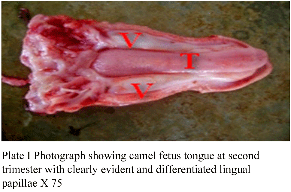 Plate I Photograph showing camel fetus tongue at second trimester with clearly evident and differentiated lingual papillae X 75 |
Table 1 The mean CVRL, sex and mean weight of the foetuses at various gestational ages |
Grossly, in all the stages of development, the tongues were observed to be elongated, with flat surfaces and rounded at the apices (Plates I-III). At first trimester, the tongues were seen as smooth muscle mass, with almost uniform width and thickness throughout the length. They were uniformly pinkish, no pigmentation and no visual evidence of lingual papillae (Plate I). At second trimester, the tongues were observed to have taken the normal shape of an adult tongue; tapering rostrally from the root to the tip or apex. The tongues were pinkish with prominent lingual papillae as evident at the posterior one-third of dorsal part of the tongue and less evident at the remaining two-thirds (Plate II). At third trimester, the tongues had developed into the typical adult tongue, with prominent lingual papillae at the tip, laterals, dorsum and the root. The tongue is relatively darkish as compared to those of first and second trimesters (Plate III). There were numerous large circumvallate papillae arranged on each side closer to one another forming two lines almost parallel to the rim of lingual torus. The shape and size of the papillae revealed remarkable differences with advancement in gestation; they were not identical or symmetrical in the two lines in the same specimen. This finding is in agreement with the previous studies of Qayyum et al., (23) on Llama; Erdunchaolu et al., (10) and Peng et al., (19) on Bacterian camel , Emura et al., (8) on sheep; abundant as in ruminants Qayyum and Beg, (22) and Prakash and Rao, (21); Chamorro et al., (4) on Horse; Kumar et al., (17) and Tadjalli and Pazhoomand, (25) on Sheep. Contrarily, single in mouse, rat and hamster by Kurtul and Atalgin, (18) and Iwasaki et al., (12).
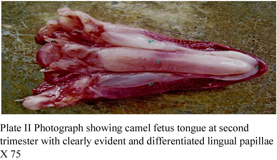 Plate II Photograph showing camel fetus tongue at second trimester with clearly evident and differentiated lingual papillae X 75 |
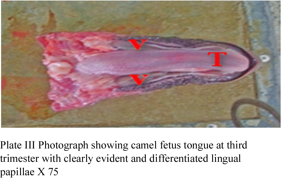 Plate III Photograph showing camel fetus tongue at third trimester with clearly evident and differentiated lingual papillae X 75 |
Biometrically, the weight of the foetuses were found to be 0.18±0.05 to 21.70±7.28kg from the first trimester to second trimester, the crown – vertebral – rump – length were found to be 15.75±4.42 to 94.00±2.83cm from the first trimester to the third trimester, weight of the head were found to be 25.05±15.17 to 1120.00±14.14g and weight of the tongues were found to be 0.79±0.22 to 116.25±11.49g from first trimester to third trimester (Table 1 and 2). The result shows that with the advancement of gestation the morphometric data were increasing progressively. This is in accordance with the finding of Qayyum et al., (22) on Llama; Erdunchaolu et al., (10) and Peng et al., (20) on Bacterian camel, Emura et al., (8) on sheep; Chamorro et al., (4) on Horse; Kumar et al., (17) and Bello et al.,(2, 3) on camel digestive tract.
Table 2 The morphometrical parameters of the tongue at the various gestational ages |
Histological observations showed that the tongue has three layers: tunica mucosa, tunica submucosa and tunica muscularis. The three layers were seen right from the first trimester, as shown in the Figure 1. The development of lingual papillae was found to be in succession; from the stages of ordinary epithelial cell linings with undifferentiated protrusions (papillae) and developing slightly into differentiated papillae, then finally to the developed papillae, with continous maturity at post-natal stage. These were seen with advancement in gestational age from first trimester through second trimester and to third trimester. Specialization of the lingual muscles also followed the same trend of development, from immature muscle fibres to mature differentiated muscle. At the first trimester, the three layers were clearly seen with slight differentiation of the epithelia into the formation of the lingual papillae (Figure 1-4). At second trimester, there was development of the three layers, with differentiation of the epithelial cells in the forming the papillae, with clear evidence of the differentiation among the papillae and the muscles of the tongue. At third trimester, the three layers were more differentiated with developed epithelial cells into differentiated lingual papillae. The maturity of the tongue can be said to continue after birth, with the formation of non-keratinised stratified squamous epithelium and the further differentiation of the muscle which were clearly evident in one-year old camel not seen at third trimester. This is in accordance with the finding of Qayyum et al., (22) on Llama; Erdunchaolu et al., (10) and Peng et al., (19) on Bacterian camel, Emura et al., (7) on sheep; Chamorro et al., (4) on Horse; Kumar et al., (17).
Figure 1 Photomicrograph of camel tongue (DORSAL) at first trimester showing Tunica mucosa (E), Tunica submucosa (S) and Tunica muscularis (M) with slight differentiation of the epithelia cells in the formation of papillae (black arrow) H&E x200 |
.png) Figure 2 Photomicrograph of camel tongue (DORSAL) at second trimester showing Tunica mucosa (E), Tunica submucosa (S) and Tunica muscularis (M) with differentiation of the epithelia cells in the formation of papillae; Circumvallate red arrow) H&E x200 |
Figure 3 Photomicrograph of camel tongue (DORSAL) at third trimester showing Tunica mucosa (E), Tunica submucosa (S) and Tunica muscularis (M) with differentiation of the epithelia cells in the formation of papillae; Circumvallate (green arrow), H&E x200 |
.png) Figure 4 Photomicrograph of camel tongue (DORSAL) at third trimester showing Tunica mucosa (E), Tunica submucosa (S) and Tunica muscularis (M) with developed non-keretinised stratified squamous epithelium (black arrow), well developed taste buds and developed differentiated skeletal muscle (M) H&E x300 |
Histological observations showed that the circumvallate papillae were generally lined with keratinised stratified squamous epithelium, which was composed by basal, spinosum, granulosum and corneum layers. Lingual glands located in the deeper parts of the papillae, and they opened into the annular groove. The surface epithelium was less keratinised compared to that of the surrounding annular pad except the peripheral parts of the papilla. The above findings coincide with those reported in Bacterian camel by Qayyum et al.,(23) and Erdunchaolu et al., (10), on Horse by Chamorro et al., (4); on cattle by Karadag et al., (13) and on deer by Adnyane et al., (1). In addition, we also observed taste buds on both sides of the secondary groove and within the epithelial lining of deep sinuses in the lateral papillary wall, which has not been reported before. In contrast, Doughbag (5) and Salehi et al. (24) found taste buds on the dorsal surface of the circumvallate papillae of the embryonic tongue. Dermal inter-digitations of variable sizes into the epidermis were observed along the whole surface. Few taste buds were observed along the medial papillary wall epithelium of the small-sized papillae. In the typical papillae, several taste buds with prominent taste pores were found along the whole length of the medial papillary wall epithelium at second trimester. However, they were concentrated at the deeper parts of the medial wall at third trimester (Figure 2 and 3). The taste buds were spindle or columnar-shaped with variability in diameter.
http://dx.doi.org/10.1111/j.1439-0264.2010.01041.x
doi:10.4172/2161-0940.1000131
http://dx.doi.org/10.4172/2161-0940.1000131
http://dx.doi.org/10.1159/000146141
http://dx.doi.org/10.1679/aohc.69.257
http://dx.doi.org/10.1046/j.1439-0264.2000.00283.x
http://dx.doi.org/10.2535/ofaj1936.77.2-3_39
http://dx.doi.org/10.2535/ofaj.85.29
http://dx.doi.org/10.1111/j.1439-0264.2001.t01-1-0317.x
http://dx.doi.org/10.1046/j.1439-0264.2001.00317.x
http://dx.doi.org/10.1111/j.1439-0264.2001.t01-1-0 317.x
http://dx.doi.org/10.1679/aohc.51.53
http://dx.doi.org/10.1002/(SICI)1097-0185(199704)247:4<528::AID-AR12>3.0.CO;2-R
http://dx.doi.org/10.3923/javaa.2010.123.130
http://dx.doi.org/10.1111/j.1439-0264.1998.tb00207.x
http://dx.doi.org/10.1016/j.smallrumres.2008.09.003
http://dx.doi.org/10.1002/cne.10551
http://dx.doi.org/10.1159/000145264
http://dx.doi.org/10.1159/000144533
http://dx.doi.org/10.3923/javaa.2010.514.518
Tadjalli, M., and R. Pazhoomand, 2004: Tongue papillae in lambs: a scanning electron microscopic study. Small Rumin. Res. 54, 157-164
http://dx.doi.org/10.1016/j.smallrumres.2003.11.005


. PDF(590KB)
. FPDF(win)
. HTML
. Online fPDF
Associated material
. Readers' comments
Other articles by authors
. A. Bello
. O. O. Alimi
. M. L. Sonfada
. M.A. Umaru
. J.E. Onu
. B.I Onyeanusi
. S.A Shehu
Related articles
. Histomorphometry
. Camel
. Circumvallate
. Papillae
. Prenatal development
Tools
. Email to a friend
. Post a comment